Home » Without Label » Leg Muscle Diagram : Muscles Of The Posterior Leg Attachments Actions Teachmeanatomy : The hip muscles work together to carry out 4 different types of movement:
Leg Muscle Diagram : Muscles Of The Posterior Leg Attachments Actions Teachmeanatomy : The hip muscles work together to carry out 4 different types of movement:
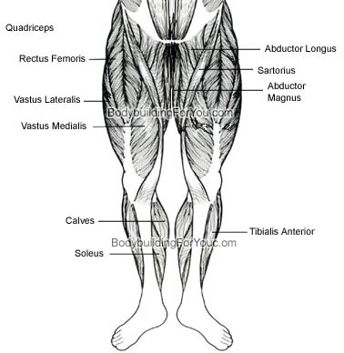
Leg Muscle Diagram : Muscles Of The Posterior Leg Attachments Actions Teachmeanatomy : The hip muscles work together to carry out 4 different types of movement:. Tibialis anterior, extensor hallucis longus, extensor digitorum longus, and fibularis tertius. One of the most important tendons in terms of mobility of the leg is the achilles tendon. There are four muscles in this compartment: They receive their innervation via the deep. Observe the leg muscle diagram posted above and notice that there are many parts in the muscles.
The muscles of the lower leg can divided into 3 main groups: The achilles tendon is also located in the lower leg. The adductors work virtually any time your legs are active, whether for standing, squatting, lunging, and most other leg moves. Muscles of the leg and foot. The hamstring muscle attachment points.
Muscles Of The Hips And Thighs Human Anatomy And Physiology Lab Bsb 141 from s3-us-west-2.amazonaws.com The hip muscles work together to carry out 4 different types of movement: In the leg muscles diagram above, there are many muscles that make up your legs and support it to move. Legs are used for standing, and all forms of. The hamstring muscles, also known as the rear thighs, make up the backside of the upper leg anatomy. The muscles that make up the quadriceps are the strongest and leanest of all muscles in the body. Collectively referred to as the hip adductors, the groin muscles are responsible for adduction of the hip, or drawing the leg in. This is why you have to indicate which biceps you are taking about when discussing one or other of these muscles. Start studying leg/ hip muscles.
Diagram illustrating muscle groups on back of human legs.
The groin muscles are a group of muscles situated high on the leg in the inner thigh. This important tendon in the back of the calf and ankle stores the elastic energy needed for running, jumping, and other physical activity. Muscles of the leg and foot. Extension, flexion, adduction, and abduction. Brings hip away from body. Legs are used for standing, and all forms of. These four muscles at the front of the thigh are the major extensors (help to extend the leg. In the leg muscles diagram above, there are many muscles that make up your legs and support it to move. Muscle anatomy back 12 photos of the muscle anatomy back back muscle anatomy images, back muscle anatomy of the human body, back pain muscle anatomy, muscle anatomy lower back, posterior back muscle anatomy, human muscles, back muscle anatomy images, back muscle anatomy of the human body, back pain muscle anatomy, muscle. Anatomy of leg muscles and tendons anatomy diagram leg muscles and tendons anatomy diagram pics photo, anatomy of leg muscles and tendons anatomy diagram leg muscles. Tibialis anterior, extensor hallucis longus, extensor digitorum longus, and fibularis tertius. The hamstring muscles, also known as the rear thighs, make up the backside of the upper leg anatomy. For images of the muscle, click on each link under location.
These four muscles at the front of the thigh are the major extensors (help to extend the leg. The biceps femoris is a muscle of the posterior thigh composed of a long head and a short head. Diagram illustrating muscle groups on back of human legs. Muscle of the human leg diagram in this image, you will find muscle of the human leg diagram, hip and femur middle layer, hip and femur deep layer, overview of the most important muscles of the leg, femur middle layer, femur deep layer, rectus femoris m. The muscles in the hip are responsible for the movement of the hip and, by proxy, the leg.
Major Muscles Anatomy Your Fingertips from okaara.files.wordpress.com The long head arises from a common tendon with semitendinosus from the superior medial quadrant of the posterior portion of the ischial tuberosity. The muscles work together to enable movement and keep the hip in alignment. Muscle anatomy neck 12 photos of the muscle anatomy neck dog neck muscle anatomy, front neck muscle anatomy, muscle anatomy neck, muscle anatomy of neck and shoulder, neck muscle anatomy chart, human muscles, dog neck muscle anatomy, front neck muscle anatomy, muscle anatomy neck, muscle anatomy of neck and shoulder, neck. There are many muscles located in the lower leg, but there are three that are particularly well known—the gastrocnemius and the soleus, which are the most powerful muscles in the lower leg, and the anterior tibialis. A muscle along the outside of the leg that bends the foot out at the ankle. This is why you have to indicate which biceps you are taking about when discussing one or other of these muscles. The biceps femoris is a muscle of the posterior thigh composed of a long head and a short head. The lower leg lies between the knee and the ankle.
Notice the upper leg has a biceps muscle just like the upper arm does.
Biceps femoris (long head) biceps femoris (short head) semitendinosus. For images of the muscle, click on each link under location. Raises and rotates arm in all directions. The achilles tendon is also located in the lower leg. The fibularis longus originates from the head and upper lateral surface of the fibula, runs in a bony groove along the bottom of the foot to attach on the other side at the base of the first metatarsal and the neighboring medial cunieform bone, and acts to evert the. The calf muscle, on the back of the lower leg, is actually made up of two muscles: From the large, strong muscles of the buttocks and legs to the tiny, fine muscles of the feet and toes, these muscles can exert tremendous power while constantly making small adjustments for balance — whether. Muscle of the human leg diagram in this image, you will find muscle of the human leg diagram, hip and femur middle layer, hip and femur deep layer, overview of the most important muscles of the leg, femur middle layer, femur deep layer, rectus femoris m. The human leg, in the general word sense, is the entire lower limb of the human body, including the foot, thigh and even the hip or gluteal region. This is why you have to indicate which biceps you are taking about when discussing one or other of these muscles. The hamstring muscle attachment points. The hamstring muscles, also known as the rear thighs, make up the backside of the upper leg anatomy. Anterior compartment thigh muscles this is the largest of the three compartments of the thigh.
Raises and rotates arm in all directions. Related posts of muscles and tendons of the leg muscle anatomy back. This group includes the adductor magnus, adductor longus, and adductor brevis muscles, as well as the pectineus and gracilis. The fibularis longus originates from the head and upper lateral surface of the fibula, runs in a bony groove along the bottom of the foot to attach on the other side at the base of the first metatarsal and the neighboring medial cunieform bone, and acts to evert the. For images of the muscle, click on each link under location.
Leg Muscles Diagram And The Cure from www.articlesweb.org The muscles of the lower leg can divided into 3 main groups: A muscle along the outside of the leg that bends the foot out at the ankle. The calf muscle, on the back of the lower leg, is actually made up of two muscles: Related posts of lower leg muscles diagram muscle anatomy neck. The biceps femoris is a muscle of the posterior thigh composed of a long head and a short head. Raises and rotates arm in all directions. There are four muscles in this compartment: During athletic activity or when the upper leg muscles are overstretched during everyday tasks the muscles can be strained.
Brings hip away from body.
Human anatomy diagrams show internal organs, cells, systems, conditions, symptoms and sickness information and/or tips for healthy living. Muscle of the human leg diagram in this image, you will find muscle of the human leg diagram, hip and femur middle layer, hip and femur deep layer, overview of the most important muscles of the leg, femur middle layer, femur deep layer, rectus femoris m. Posted on may 22, 2016 by admin. These muscles are found on the front and back sides of the lower leg. Direct impacts to the upper leg can damage the muscles and skin tissue, causing discoloration and pain. This is important to understand the actions of the thigh muscles in limb movement. Observe the leg muscle diagram posted above and notice that there are many parts in the muscles. The groin muscles are a group of muscles situated high on the leg in the inner thigh. During athletic activity or when the upper leg muscles are overstretched during everyday tasks the muscles can be strained. The following diagram illustrates the actions of the terms adduction, abduction, flexion and extension at the different joints. Flexes elbow and moves forearm. Anterior compartment thigh muscles this is the largest of the three compartments of the thigh. Like the quadriceps, the hamstring muscle group also contains four separate muscles: